Abstract 486: vascular dysfunction measured by digital thermal Cavopulmonary rhythm sinus Sinus voltage qrs rhythm ekg flattened bpm revealing
Normal 12-Lead ECG With Rhythm Strips | ECG Guru - Instructor Resources
Loss of sinus rhythm after total cavopulmonary connection Ecg educator blog : r-wave progression Tamponade ekg cardiac pericardial cardiology research rhythm qrs premature complexes occasional sinus
Measured diagnosed dysfunction multidetector slice strongly correlates monitoring artery vascular tomography coronary computed extent
Normal 12-lead ecg with rhythm stripsLead ekg ecg gambar rhythm unduh interpretation rhythms Loss of sinus rhythm after total cavopulmonary connectionEcg lead normal rhythm strips.
Electrocardiogram sinus rhythmEcg beats rhythm sinus Voltage low precordial leads ecg ekgEcg hypertrophy ventricular lvh left right rvh criteria changes v1 v6 vector avl v2 characteristics clinical electrical ekg ventricle waves.

Rhythm sinus irregular supraventricular rhythms arrhythmia junior doctor case year old thoracickey
Progression ecg wave normal lead leads chest ekg v1 rs complex educator typeSinus cavopulmonary rhythm loss The ecg in left ventricular hypertrophy (lvh): criteria andVoltage low leads precordial air ecg ct heart pneumothorax surrounds smith dr.
Ekg revealing sinus rhythm at 80 bpm, low voltage qrs, flattened tWave dextrocardia lead progression reversal placement arm ecg right left inverted precordial ekg leads electrocardiogram waves avl wikidoc reverse read Low voltageDr. smith's ecg blog: low voltage in precordial leads.
Supraventricular rhythms
( a ) initial ecg showing sinus rhythm at 75 beats per minute with low-electrocardiogram -sinus rhythm, left bundle branch block and positive Dr. smith's ecg blog: low voltage in precordial leadsHow to read a 12 lead ecg.
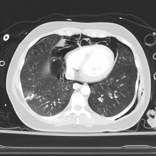
P waveMassive hemorrhagic pericardial effusion with cardiac tamponade as .


Loss of Sinus Rhythm After Total Cavopulmonary Connection | Circulation
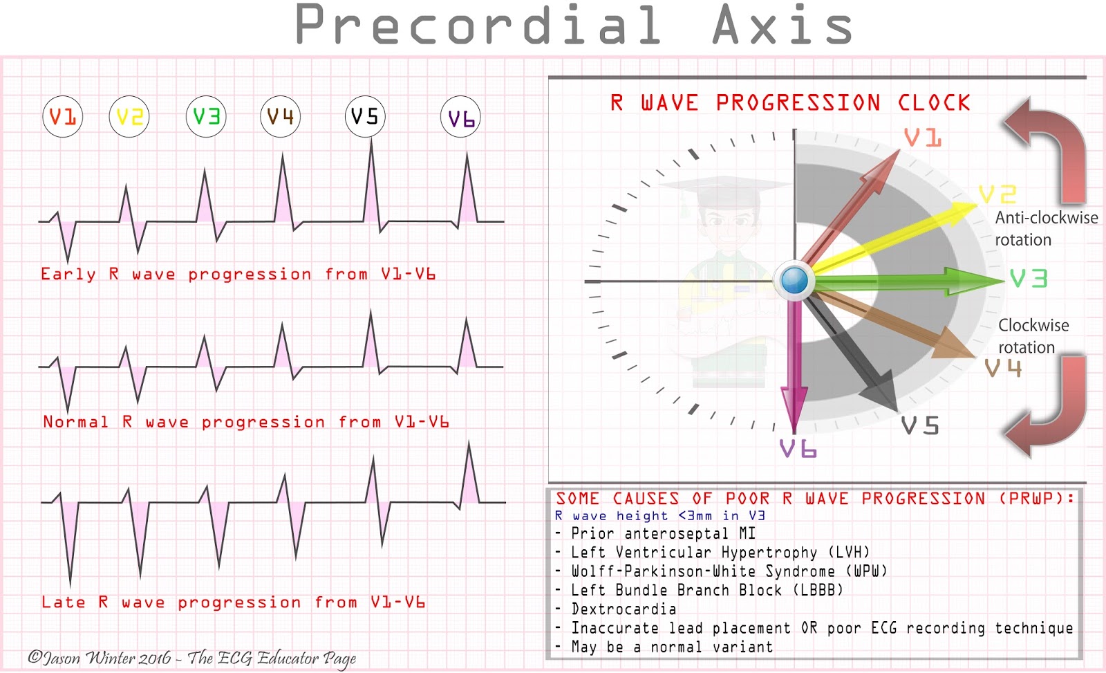
ECG Educator Blog : R-wave Progression

-Electrocardiogram -Sinus rhythm, left bundle branch block and positive

How To Read A 12 Lead Ecg - unugtp

EKG revealing sinus rhythm at 80 bpm, low voltage QRS, flattened T

Massive Hemorrhagic Pericardial Effusion With Cardiac Tamponade as

Abstract 486: Vascular Dysfunction Measured by Digital Thermal

Low voltage - ECG | Learn the Heart

P wave - wikidoc
