Exposure technique factors Kvp factors rays ray technique radiography exposure ppt powerpoint presentation does produced quantity higher energy quality slideserve Kvp contrast
PPT - [Radiography] Technique - Exposure Factors PowerPoint
High kvp chest radiograph: (a) original image and (b) processed image Radiology tutorials: x-rays Kv ray radiography tube voltage effect radiology kvp contrast medical changing upstate film
Radiographic exposure technique
Exposure kvp radiation receptor increasing increases penetrating power factors technique radiology figure radiologykeyEffect of changing x-ray tube voltage (kv) Technique radiographic factors kvp exposure ray radiology penetration beam quality digital absorption exit intensities figure radiologykey relationshipKvp radiograph.
Dual energy radiography acquisition and processingKvp energy dual richard imaging bone Kvp and contrastKvp cerebral versus perfusion regional powerpoint ajnr.

Samuel richard
Radiography radiology dual energy acquisition soft processing rsna upstate bone above indexRadiographic exposure technique Using 80 kvp versus 120 kvp in perfusion ct measurement of regionalRadiographic exposure technique kvp density factors radiology increasing radiation radiographs increases range relationship figure radiologykey receptor important.
Radiology rays properties animated .

![PPT - [Radiography] Technique - Exposure Factors PowerPoint](https://i2.wp.com/image.slideserve.com/546181/slide2-l.jpg)
PPT - [Radiography] Technique - Exposure Factors PowerPoint
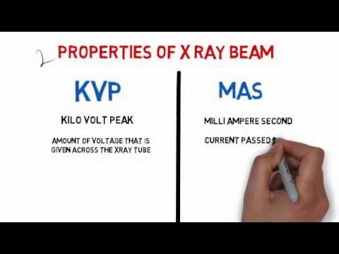
Radiology Tutorials: X-Rays - Properties of X Rays: (Medical Animated

Samuel Richard

Dual Energy Radiography Acquisition and Processing | Radiology |SUNY

Radiographic Exposure Technique | Radiology Key
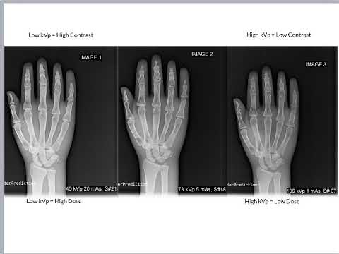
kVp and Contrast - YouTube

Exposure Technique Factors | Radiology Key

Using 80 kVp versus 120 kVp in Perfusion CT Measurement of Regional

High kVp chest radiograph: (a) original image and (b) processed image
